First find the cause
Assessment looks at symptoms, examination, thyroid blood tests and ultrasound. CT may be added if the gland is very large or extends behind the breastbone.
 Contact
A patient-friendly guide to goiter, Graves’ disease, the tests that come first, and when thyroid artery embolization may be discussed as one option for selected people.
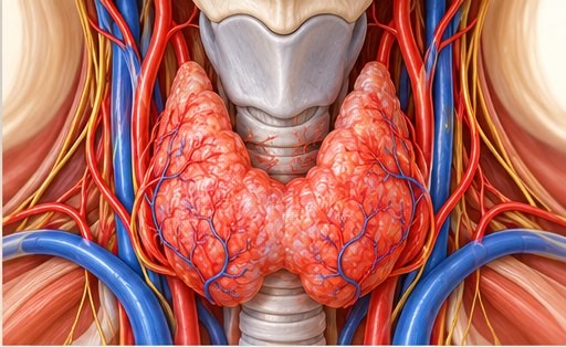
Illustrations are AI-generated for education; they are not patient images, diagnostic scans or a guide to performing a procedure.
Thyroid artery embolization (TAE) is an image-guided procedure. A doctor guides a thin catheter from an artery in the wrist or groin to selected small arteries that feed the thyroid. Tiny particles are released into those thyroid-feeding branches to reduce blood flow to the treated part of the gland.
The target is a thyroid artery, such as a superior or inferior thyroid artery branch. The main carotid arteries that carry blood to the brain are not treatment targets. The route and arteries considered depend on each person’s scans and anatomy.
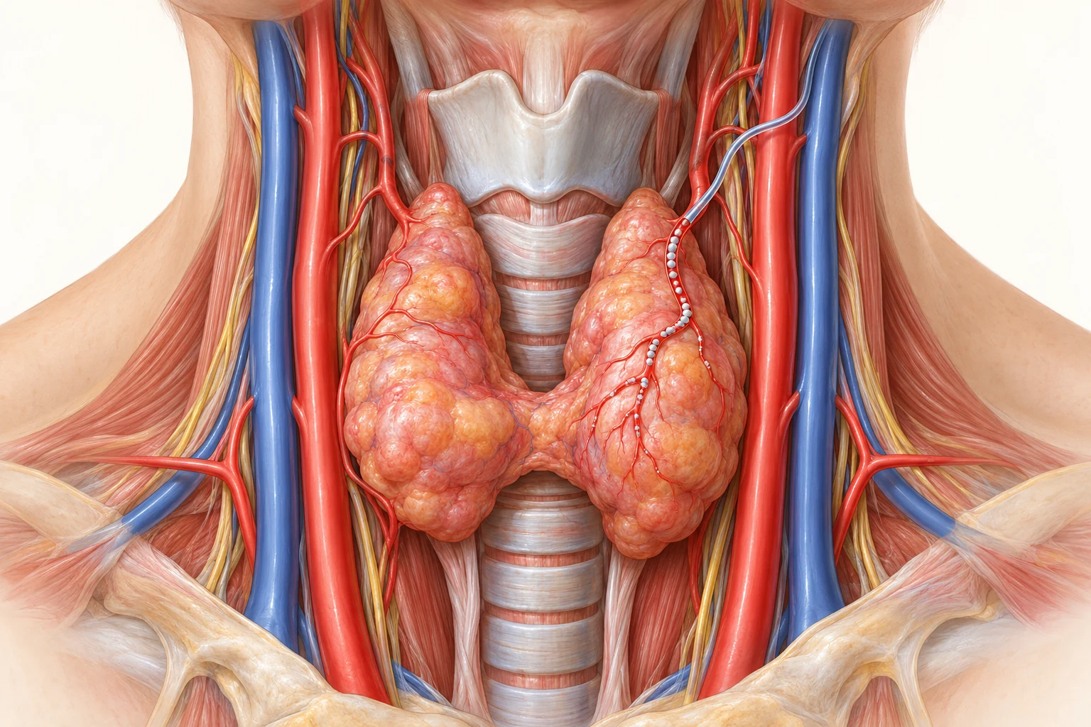
A goiter simply means that the thyroid gland is larger than usual. It can be smooth and evenly enlarged or contain one or more nodules. A goiter may produce normal, high or low thyroid hormone levels. Some people notice a neck lump, pressure, swallowing or breathing difficulty; others have no symptoms.
Assessment looks at symptoms, examination, thyroid blood tests and ultrasound. CT may be added if the gland is very large or extends behind the breastbone.
Depending on the cause, choices may include monitoring, medicines, radioactive iodine, surgery or—in selected benign nodules—needle ablation. Each has different benefits and trade-offs.
For a person with a confirmed benign goiter who is not a suitable surgical candidate, or who declines surgery after counselling, a specialist team may discuss whether TAE is reasonable alongside other available choices.
Graves’ disease is an autoimmune condition. The immune system stimulates the thyroid to make too much hormone. Symptoms can include a racing heart, tremor, heat intolerance, weight change, tiredness and sometimes eye symptoms. Blood tests and, when appropriate, thyroid antibody tests help clinicians confirm the diagnosis.
Established treatment choices can include antithyroid medicines, radioactive iodine and thyroid surgery. TAE has also been studied in selected people, but the published experience is limited and does not establish it as the right choice for every person with Graves’ disease. An endocrinologist should guide care, with thyroid surgery and interventional radiology input when TAE is being considered.
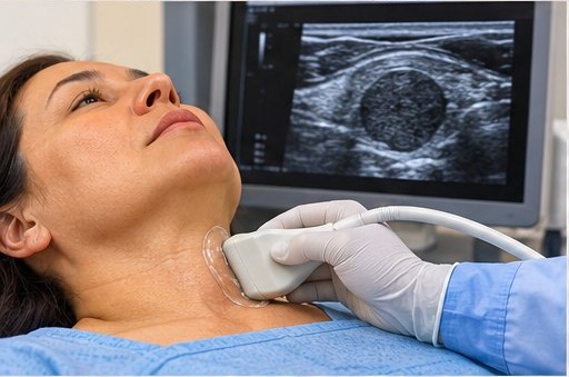
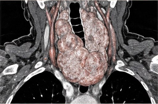
Ultrasound is usually the first imaging test. For a large goiter or one that may extend into the chest, CT can show its full size, position and relationship to nearby structures.
Tests help confirm what is causing the thyroid problem, how large the gland is, and whether a benign diagnosis is secure. Your clinician decides which investigations are appropriate for you.
Ultrasound examines the gland and nodules, measures their size, and identifies any features that need further assessment.
For a large goiter or possible extension into the chest, CT can clarify the gland’s volume, position and effect on nearby structures. The team decides whether contrast is appropriate.
Tests commonly include TSH and thyroid hormone levels. Antibody tests may help assess Graves’ disease. Other blood tests check general health and help plan a procedure if one is considered.
If a nodule’s ultrasound appearance or history indicates it, a needle sample (FNAC) may be needed. Suspicious or uncertain results need the appropriate diagnostic work-up; they should not simply be treated as benign goiter.
An endocrinologist and thyroid surgeon explain relevant options such as medicines, radioactive iodine, monitoring or surgery—and why an option may not fit your situation.
Only after diagnosis and review, the team may discuss TAE for selected people with benign disease when surgery is not suitable or is declined after informed discussion. The potential benefits, uncertainty and risks are part of consent.
The details depend on the person’s arteries, treatment plan and the hospital’s practice. These steps explain the experience in broad terms, not how to perform the procedure.
A small tube may be placed in an artery at the wrist (radial access) or groin (femoral access). The team chooses the route after reviewing your anatomy and treatment plan.
X-ray imaging and contrast help the interventional radiologist guide a thin catheter towards the arteries supplying the thyroid. The catheter is directed into selected thyroid artery branches.
Small embolic particles are delivered into selected thyroid-feeding branches. The main carotid artery is not the treatment target. A final image check is used before the catheter and access tube are removed.
You are monitored throughout. Local anaesthetic is used at the access site; some centres may also use sedation or other comfort measures. You may feel pressure at the wrist or groin, warmth when contrast is injected, or temporary neck discomfort. Tell the team straight away if you feel pain, nausea, breathlessness or anything that worries you.
The team checks the access site and observes you for a period set by your clinicians. You may have temporary neck soreness, tenderness or a flu-like feeling. Follow the centre’s instructions about medicines, activity, wound care and follow-up blood tests and imaging. Contact the care team if symptoms worsen.
Wrist and groin access are both used in published reports. The actual route, medicines, contrast and observation plan are individualized.
The right approach depends on the diagnosis, symptoms, test results and your preferences. Your care team can explain which options are appropriate for you.
Follow-up may be reasonable when symptoms and test findings allow it.
Medicines may help control thyroid hormone levels or treat Graves’ disease.
This may be an option for selected thyroid conditions after specialist assessment.
Surgery may be advised for some goiters, especially when they cause pressure or the diagnosis needs clarification.
Needle ablation may suit selected benign thyroid nodules. It is different from thyroid artery embolization.
What is causing my goiter or Graves’ disease? What do my ultrasound, CT, blood tests and any needle sample show?
Which established treatments are suitable for me? If surgery is not suitable or I do not want it, what other options can I consider?
Why might TAE fit my case? What benefit is realistic, what is still uncertain, and how many similar cases has this team treated?
What are this centre’s complication rates? How will you protect nearby arteries and nerves, monitor my thyroid tests, and follow me afterward?
These links open the published papers or professional statements. Some are written for clinicians, but the short notes here explain what each one may help you understand.
This guide is for general education. It is not a diagnosis or personal treatment recommendation.
Bring your ultrasound, blood-test and biopsy reports if available. The appropriate next step depends on your individual assessment.
Phone: +91 70348-39446 · Email: veindoctoraju@gmail.com